Computed tomography
Definition
Computed tomography (CT), formerly referred to as computerized axial tomography (CAT), is a common diagnostic imaging procedure that uses x rays to generate images (slices) of the anatomy.
Purpose
Computed tomography (CT) is an x-ray imaging procedure used for a variety of clinical applications. CT is used for spine and head imaging, gastrointestinal imaging, vascular imaging (e.g., detection of blood clots), cancer staging and radiotherapy treatment planning, screening for cancers and heart disease, rapid imaging of trauma, imaging of musculoskeletal disorders, detection of signs of infectious disease, and guidance of certain interventional procedures (e.g., biopsies). CT is the preferred imaging exam for diagnosing several types of cancers, and along with the chest x ray, CT is the most commonly performed procedure for imaging the chest. CT is also used to perform noninvasive angiographic imaging to assess the large blood vessels.
CT may be performed on newborns, infants, children, and adolescents. In children, CT is most frequently used in the hospital emergency department to evaluate the effects of trauma, especially to the head, face, brain, and spine, and to diagnose or rule out appendicitis and other abdominal disorders because a scan can be completed in less than 20 seconds. Chest CT examinations are used to assess complications from infectious diseases, such as pneumonia and tuberculosis , inflammation of the airways, and birth defects. CT scans of the pelvic area are used to image ovarian cysts and tumors, bladder abnormalities, urinary tract stones, kidney disease, and bone disease. Head CT scans are used to examine the brain and sinuses. For children with cancer, CT is used to assist in treatment planning and to monitor cancer progression and response to treatment. For children requiring complex surgeries (e.g., brain, spine), CT is often used to produce images of the anatomy that help surgeons plan the surgery. Newer CT scanners, called multislice or multidetector CT, are used to rapidly image newborns to assess congenital heart defects.
Description
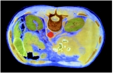
CT is performed using a specialized scanner, an x-ray system, a patient table, and a computer workstation. The CT scanner is shaped like a large square with a hole in the center or round like a doughnut. X rays are produced in the form of a beam that rotates around the patient. During a CT scan, the patient table is moved through the center hole as x-ray beams pass through the patient's body. The x rays are converted into a series of black-and-white images, each of which represents a "slice" of the anatomy.
CT scans are conducted by a technologist with specialized training in x rays and CT imaging. During scanning, the technologist operates the CT scanner using a computer located in an adjacent room. Because movement during the scan can cause inaccurate images, the technologist instructs the patient via an intercom system to hold their breath and not move during the x rays. The scan itself may only take five to 15 minutes, but total examination time may be up to 30 minutes, since the patient must be prepped and positioned. Abdominal CT scans usually require that the child drink a solution that contains a dye, called oral contrast, that shows up on the CT images to help better define internal organs. For pelvic scans, contrast material may be delivered via the rectum. Some CT scans also require the injection of contrast material into the vein to help define the blood vessels and surrounding tissue.
The images from CT examination are called slices because they are acquired in very small (millimeter-size) sections of the body. The image slices are displayed on a computer monitor for viewing or printed as a film. A radiologist interprets the x-ray images produced during the CT examination. For emergency CT scans, images are interpreted immediately so that the child can be treated as soon as possible. For non-urgent outpatient CT scans, the radiologist interprets the images and sends a report to the referring physician within a few days.
For emergency situations, CT scans are performed in a hospital radiology department in conjunction with
Precautions
CT scans expose the child to radiation, and overuse of CT scanning has received attention from organizations that regulate medical radiation exposure. Although no side effects have been linked to radiation exposure from CT imaging, the Food and Drug Administration has issued guidance to physicians regarding levels of radiation during pediatric CT examinations. New CT scanners have preset imaging features that allow scanning at the lowest radiation dose for the child's weight and age.
Oral contrast may be unpleasant tasting, although chocolate, vanilla, and fruit flavors may be available. Injected contrast can cause sensations of heat or cold through the body. Some children may have allergic reactions to the contrast material, although severe reactions are rare. Parents should inform CT staff if their child has ever had a reaction to any medication, contrast material, or anesthesia. Because contrast material may contain iodine, sensitivity to contrast material may occur if the child has other allergies to iodine or seafood, and CT staff should be informed if the child has such allergies. Also, because CT contrast material can affect kidney function, parents should notify CT staff if their child has kidney disease.
Preparation
Abdominal CT examinations usually require fasting for at least 12 hours before the scan. If the intestines will also be imaged, a laxative before the scan is required. Parents should alert CT staff if children are diabetic and taking insulin, since hypoglycemia can occur with missed meals.
Before the CT scan, the patient has to change into a hospital gown. When oral contrast is necessary, patients need to arrive at least one hour before the scan to drink the contrast solution. During the scan, the child is asked to lie on the CT table. Positioning devices, such as head cradles or knee rests, may be used. For very young or very active children, foam or Velcro restraints may be used to minimize movement during imaging. Or sedation may be used if children cannot remain still. After positioning the child, the technologist inserts an intravenous catheter to inject contrast material.
CT scanners may frighten young children, so prior to the imaging examination, the basic procedure should be explained to help reduce fear . Some radiology departments offer special patient education booklets for children that help explain imaging procedures.
Aftercare
No special aftercare is required following CT scans, unless sedation or general anesthesia was used during the scan. In these cases, children are required to remain in a supervised recovery area for an hour or more following the procedure to be monitored for reactions to anesthesia. If injected contrast material is used, some minor first aid (small bandage, pain relief) for the injection site may be necessary.
Risks
Radiation exposure is a risk during CT examinations. However, the radiation from a CT scan is usually less than that from regular x rays, and the benefits of the examination far outweigh the minor radiation dose received during the scan.
Some children may have reactions to anesthesia or sedation, including headaches, shivering, or vomiting . Rarely, severe anaphylactic reactions can occur that require emergency treatment.
Parental concerns
Younger children may be frightened of the CT scanner, and a parent or other family member may be required to be present in the scanning room. To help alleviate fear, taking the child into the CT room to see the equipment prior to the procedure may be helpful. To reduce risk of radiation exposure, anyone remaining in the scanning room during x-ray delivery will have to wear a lead apron on shield.
KEY TERMS
Anaphylaxis —Also called anaphylactic shock; a severe allergic reaction characterized by airway constriction, tissue swelling, and lowered blood pressure.
Radiologist —A medical doctor specially trained in radiology, the branch of medicine concerned with radioactive substances and their use for the diagnosis and treatment of disease.
Resources
BOOKS
Margolis, Simeon, et al. The Johns Hopkins Consumer Guide to Medical Tests: What You Can Expect, How You Should Prepare, What Your Results Mean. New York: Rebus Inc., 2002.
Medical Tests: A Practical Guide to Common Tests. Boston, MA: Harvard Health Publications, 2004.
Segen, J. C., et al. The Patient's Guide to Medical Tests: Everything You Need to Know about the Tests Your Doctor Prescribes. New York: Facts on File, 2002.
Shannon, Joyce Brennfleck. Medical Tests Sourcebook: Basic Consumer Health Information about Medical Tests. Detroit, MI: Omnigraphics, 2004.
PERIODICALS
Harvey, D. "Evaluating Pediatric Trauma: Imaging vs. Lab Tests." Radiology Today 5 (August 2, 2004): 14–16.
ORGANIZATIONS
American College of Emergency Physicians. 2121 K St., NW, Suite 325, Washington, DC 20037. Web site: http://www.acep.org.
American College of Radiology. 1891 Preston White Dr., Reston, VA 20191. Web site: http://www.acr.org.
Radiological Society of North America. 820 Jorie Blvd., Oak Brook, IL 60523–2251. Web site: http://www.rsna.org/.
WEB SITES
"CT Scan." Emedicine , November 1, 2004. Available online at http://www.emedicinehealth.com/Articles/11618-1.asp (accessed December 21, 2004).
"Pediatric CT (Computerized Tomography)." Radiology Info: The Radiology Information Source for Patients. Available online at http://www.radiologyinfo.com/content/pediact.htm (accessed December 21, 2004).
Jennifer Sisk, MA