Magnetic resonance imaging
Definition
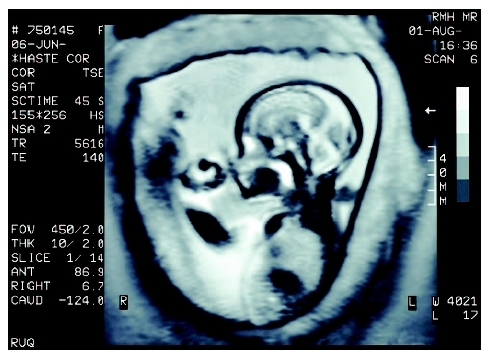
Magnetic resonance imaging (MRI) is a diagnostic imaging procedure that uses radio waves, a magnetic field, and a computer to generate images of the anatomy.
Purpose
MRI is used to visualize the body to assist doctors in their efforts to diagnose certain diseases or conditions and to evaluate injuries. For pediatric imaging, MRI is used for a variety of purposes, including the following:
- diagnosing diseases of the central nervous system, including the brain and spine
- detecting musculoskeletal disorders and injuries
- identifying complications of infectious diseases, such as those associated with Lyme disease or acquired immunodeficiency syndrome (AIDS)
- imaging the cardiovascular system
- detecting congenital heart defects in neonates
- determining the stage of certain types of cancer
- evaluating bone marrow disease
- assessing blood vessels in the brain for stroke and other abnormalities
- assisting in the planning of surgery and cancer treatment
- evaluating the urinary tract
MRI provides images with excellent contrast that allow clinicians to clearly see details of soft tissue, bone, joints, and ligaments. MRI angiography is an imaging technique used to evaluate the blood vessels, for example, to detect aneurysms or cardiovascular problems. Because MRI does not use ionizing radiation to produce images, like x ray and CT, it is often the examination of choice for pediatric imaging and for imaging the male and female reproductive systems, pelvis and hips, and urinary tract and bladder.
MRI can also be used to evaluate brain function for assessing language, senses, neurologic disorders, and pain . This technique, called functional MRI, involves rapid imaging to display changes in the brain's blood flow in response to tasks or visual and auditory stimuli. Functional MRI is being researched to image neurologic disorders, such as attention deficit hyperactivity disorder (ADHD), delayed cognitive development , and epilepsy.
MRI spectroscopy is another emerging imaging technique for evaluating pediatric brain disorders. In MRI spectroscopy, chemicals in the brain are measured and brain tissue is imaged. This technique is being investigated to evaluate traumatic brain injury, speech delay, creatine deficiency syndromes, and mood disorders in young children.
Interventional and intraoperative MRI is another developing field that involves performing interventional procedures, primarily brain surgeries, using a specially designed MRI unit in an operating room.
Description
MRI is performed using a specialized scanner, a patient table, systems that generate radio waves and magnetic fields, and a computer workstation. The scanner, which is usually shaped like a large rectangle with a hole in the center, contains the systems that generate the magnetic field. A motorized and computer-controlled patient table moves into the scanner's center hole during the scan. A technologist operates the MRI scanner from an adjacent control room that contains a computer system and an intercom system for communicating with the patient during the scan.
In most MRI scanners, the patient opening is like a long tube, and some patients may become claustrophobic. To be more patient-friendly, different types of MRI scanners have been developed. Newer MRI scanners have shorter patient openings that allows the patient's head to remain outside the machine during body scans. Open MRI scanners are available with columns and open sides to alleviate claustrophobia.
Depending on the body area being scanned, special body coils may be used to enhance the images. These coils are foam and plastic braces or wraparound pads that are placed on the body part being imaged. For head imaging, the coil may be shaped like a head or neck rest.
Children undergoing an MRI scan are appropriately positioned on the patient table by the technologist. For some scans, an injected contrast material may be used and is administered using an intravenous catheter. Once the patient is positioned, the technologist goes to an adjacent control room to operate the scanner. The technologist uses an intercom system to instruct the child to hold their breath or remain still at certain times during the scan. Scans range from 30 minutes to 90 minutes, depending on the type of scan. When the MRI machine is scanning, the child hears loud clanging and whirring noises. To alleviate fear or stress related to hearing this noise and being in the small scanning tube, the child may be offered earplugs or specially designed head phones for listening to music. Centers that specialize in pediatric imaging often also have special video goggles so that the child can watch a cartoon or movie during the scan. For infants, neonatal noise guards—special padded ear shields—are available.
MRI scans are performed in a hospital radiology department for inpatients and emergency cases. For scans requested by a physician, the MRI examination can be performed in the hospital radiology department on an outpatient basis or in an imaging center. Hospitals that do not have their own MRI systems may schedule MRI scans by contracting with a company that brings an MRI scanner in a specially designed mobile trailer. Mobile MRI services are frequently used in rural areas. For some conditions, such as orthopedic disorders or injuries, an MRI may be performed in a physician's office using a small MRI unit called an extremity MRI scanner. These scanners are designed to image only the joints or the head. During this type of scan, only the body part to be scanned is placed in the smaller scanner while the patient lies on a couch or sits in a chair.
The images from an MRI examination are called slices, because they are acquired in very small (millimeter-size) sections of the body. The image slices are displayed on a computer monitor for viewing or printed as a film. A specialist called a radiologist interprets the images produced during the MRI examination. For emergency scans, images are interpreted immediately so that the child can be treated quickly. For non-urgent outpatient MRI scans, the radiologist interprets the images and sends a report to the referring physician within a few days.
Precautions
MRI is a safe procedure that does not involve radiation. However, the magnetic field generated during an MRI examination is so strong that metal objects or objects with metal in them, such as jewelry, eyeglasses , oxygen canisters, and even wheelchairs, will be pulled toward the machine. Therefore, MRI staff must take special precautions to ensure that no metallic objects enter the MRI suite. MRI technologists inspect patient clothing and accessories to make sure there are no metals on them during the scan.
Preparation
Prior to any MRI scan, patients are required to remove all metal objects and remove any clothing with metal on them (zippers, snaps). In most cases, parents have to complete a survey regarding their child's past surgical procedures and medical history to indicate whether the child has any metallic implants. Metallic implants include artificial joints, pacemakers, aneurysm clips, metal plates, pins or screws, and surgical staples. Children with metallic implants are likely to undergo a computed tomography (CT) examination instead of an MRI.
Unlike CT, no fasting or laxatives are required prior to an MRI scan. Only one type of MRI scan, called a magnetic resonance cholangiopancreatography (MRCP), which scans the bile ducts, requires that the child not eat or drink anything for two to three hours prior to the scan.
During the examination, the child must lie still. The MRI scanner does make loud noises throughout the examination, which can be frightening for some children. Before the examination, the procedure should be explained to the child, and it should be emphasized that the examination is painless. Most facilities have specially designed music systems so that patients can wear headsets and listen to music during the scan; some facilities even have special video goggles so children can watch a cartoon or movie during the scan.
KEY TERMS
Anaphylaxis —Also called anaphylactic shock; a severe allergic reaction characterized by airway constriction, tissue swelling, and lowered blood pressure.
Cholangiopancreatography —An examination of the bile ducts and pancreas.
Claustrophobia —Fear of small, enclosed spaces.
Computed tomography (CT) —An imaging technique in which cross-sectional x rays of the body are compiled to create a three-dimensional image of the body's internal structures; also called computed axial tomography.
Intravenous —Into a vein; a needle is inserted into a vein in the back of the hand, inside the elbow, or some other location on the body. Fluids, nutrients, and drugs can be injected. Commonly called IV.
Radiography —Examination of any part of the body through the use of x rays. The process produces an image of shadows and contrasts on film.
Radiologist —A medical doctor specially trained in radiology, the branch of medicine concerned with radioactive substances and their use for the diagnosis and treatment of disease.
Aftercare
No special aftercare is required following MRI scans, unless sedation or general anesthesia was used during the scan. Then children are required to remain in a supervised recovery area for an hour or more following the procedure to monitor for reactions to anesthesia. If injected contrast material is used, some minor first aid (small bandage, pain relief) for the injection site may be necessary.
Risks
MRIs present no radiation exposure. Magnetic fields used in MRI have no side effects for the patient. The contrast material used in MRI contains a material called gadolinium, that is much less likely to cause severe anaphylactic (allergic) reactions than the iodinated material used for CT scans.
Because the MRI examination is long and the patient opening in the machine is small, some children and adolescents may feel claustrophobic. Light sedation or relaxants may be administered, or an MRI scanner with a more open design may be used. For younger infants and children that require sedation or anesthesia to complete the examination, reactions to the anesthesia are possible, including headaches and vomiting .
Parental concerns
Younger children may be frightened of the MRI scanner, and a parent or other family member may be required to be present in the scanning room. To help alleviate fear, taking the child into the MRI room to see the equipment prior to the procedure may be helpful. Anyone remaining in the scanning room during the MRI examination must remove any metal objects, including jewelry and eyeglasses.
Resources
BOOKS
Medical Tests: A Practical Guide to Common Tests. Boston, MA: Harvard Health Publications, 2004.
PERIODICALS
Harvey, D. "Evaluating Pediatric Trauma: Imaging vs. Lab Tests." Radiology Today 5 (August 2, 2004): 14–16.
Panigrahy, A., et al. "Advances in Magnetic Resonance Imaging of Pediatric Congenital Heart Disease." Applied Radiology. Supplement (June 2002): 103–11.
Surface, D. "MRI Spectroscopy and Pediatric Brain Disorders." Radiology Today 4 (August 4, 2003): 6–8.
ORGANIZATIONS
American College of Radiology. 1891 Preston White Dr., Reston, VA 20190. Web site: http://www.acr.org.
Radiological Society of North America. 820 Jorie Blvd., Oak Brook, IL 60523–2251. Web site: http://www.rsna.org.
WEB SITES
"Magnetic Resonance Imaging (MRI)." eMedicine Consumer Health , July 13, 2004. Available online at http://www.emedicinehealth.com/Articles/6622-1.asp (accessed November 29, 2004).
"MR Imaging (MRI)—Body." Radiology Info: The Radiology Information Source for Patients , August 2004. Available online at http://www.radiologyinfo.com/content/mr%5Fof%5Fthe%5Fbody.htm (accessed November 29, 2004).
Jennifer Sisk, MA
Comment about this article, ask questions, or add new information about this topic: