Hernia
Definition
A hernia is the protrusion of an organ through the structure or muscle that usually contains it.
Description
There are many different types of hernias in children. The most common are direct inguinal hernias, indirect inguinal hernias, and umbilical hernias. A direct inguinal hernia occurs when a small section of bowel herniates, or protrudes, through the groin muscle. Indirect inguinal hernia occurs when part of the bowel protrudes through the muscles of the groin into a sac left over from fetal development. An umbilical hernia occurs when a portion of the bowel protrudes through a small defect in the abdominal wall muscle near where the umbilical cord attaches to the baby's abdomen. More serious defects involving herniation of abdominal contents outside the infant's body are omphalocele and gastroschisis. These are not a result of an organ protruding through weakened muscle tissue but rather are a result of a much larger defect of the muscles of the abdomen that causes the internal organs to develop outside the body. Omphalocele and gastroschisis are considered abdominal wall defects and are not called hernias.
While an umbilical hernia usually resolves spontaneously as the abdominal muscles grow and requires no further treatment, in children with direct and indirect inguinal hernia, surgery is almost always required to prevent the herniated bowel from becoming incarcerated or strangulated. When an inguinal hernia is incarcerated, the bowel becomes swollen and trapped outside the body. If the hernia remains incarcerated for too long, strangulation can occur. In strangulation, the blood supply to the section of bowel that has herniated is cut off, and the tissue begins to die. When this happens, the intestines cannot function properly and are said to be obstructed. If the bowel perforates, or develops a hole in it, emergency surgery is required to repair the intestine and prevent infection.
A more severe, but less common, hernia is a diaphragmatic hernia. This occurs inside the body when the diaphragm, the large muscle that separates the abdominal cavity from the chest cavity, fails to develop fully. In children with diaphragmatic hernia, the contents of the abdomen protrude into the chest cavity. These children may have difficulty breathing. During fetal development the presence of abdominal organs in the fetal chest cavity prevents the lungs from growing normally. A diaphragmatic hernia can occur as an isolated defect or as part of a more complex syndrome. Children with diaphragmatic hernias are usually very ill and require immediate treatment after birth. Some of these children have other defects such as cardiac anomalies, chromosomal abnormalities, kidney and genital anomalies, and neural tube defects, such as spina bifida .
Demographics
Estimates of the true incidence of inguinal hernias vary, but they may affect 1–5 percent of all births in the United States. International rates appear to be similar. Males are more than seven times more likely to have an inguinal hernia than females, and premature infants are more likely than full term infants to have inguinal hernias and to have incarcerated hernias. While inguinal hernias seem to affect all racial groups at the same rate, umbilical hernias occur more frequently in African Americans.
Diaphragmatic hernias occur in approximately one in every 3,000 births. These hernias do not seem to affect any race or nationality more than another.
Causes and symptoms
A direct inguinal hernia is caused when the muscles of the floor of the groin area are weak and allow the bowel to press through. An indirect inguinal hernia is caused when remnants of early fetal genital development stay within the body after this development is complete. In early fetal development male and female genitalia are identical. At around the seventh week of gestation, the gonads (sex organs) begin to change, or differentiate, into the characteristic genitalia of males and females. Males develop testes, and females develop ovaries. During this process, in some fetuses, a small sac may form near the genitalia. Most often the opening to this sac, called the processus vaginalis, closes. However, in children with inguinal hernia, this sac remains patent, or open, becoming a container into which bowels may be herniated.
The main symptom of inguinal hernias (both direct and indirect) in infants is an obvious bulge in the groin in the inguinoscrotal region (near the scrotum) in boys and in the inguinolabial (near the labia) in girls. The bulge may or may not be painful. It will usually appear after straining or crying and then disappear after a period of time. If the hernia has incarcerated, the infant will be in obvious pain , appearing fussy, crying, and refusing to eat. The skin over the hernia may be discolored and swollen.
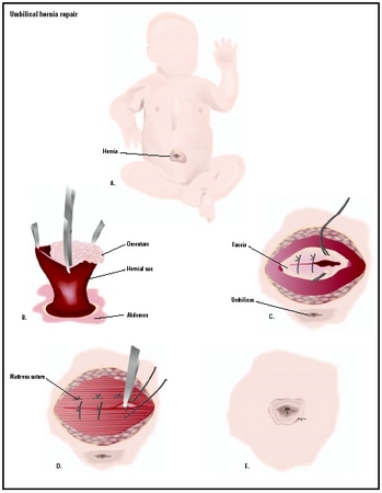
Umbilical hernia is caused by a small defect in the muscles of the abdominal wall. These hernias are usually small and have no symptoms other than a small protrusion near the base of the umbilical cord.
Like inguinal hernias, diaphragmatic hernias are caused early in fetal development. The structures that form the diaphragm do not properly form, allowing the contents of the lower abdomen to migrate up near the heart and lungs. The increased pressure these organs place on the lungs causes the lungs to remain small and underdeveloped. When the infant is born and must breathe air, the lungs are not able to work properly.
Children with diaphragmatic hernia have the following symptoms immediately after birth: breathing difficulty, a bluish skin color (cyanosis), rapid breathing, rapid heat rate, and asymmetrical chests—one side is not the same size as the other. These infants are often critically ill and are be placed on a ventilator—a machine to help them breath. Because the lungs have not had enough room to grow and are small, doctors must stabilize the baby's breathing before the hernia can be repaired.
When to call the doctor
If a small child, especially an infant, has a bulge in the abdominal or groin area, the child's pediatrician should be consulted. If the child is in severe pain, and the skin is discolored or swollen, medical help should be sought immediately.
Diagnosis
Umbilical and inguinal hernias are diagnosed by physical examination. For some children with inguinal hernia, a laparoscopic examination may be performed. A laparoscopy is an exploratory surgical procedure in which the doctor makes an incision and inserts a small tube connected to a camera to view the herniated area. This procedure is used most often in patients who have already had one hernia repair to see if the hernia has returned in a new location.
Diaphragmatic hernia may be diagnosed while the fetus is still in the womb using prenatal ultrasonography. After birth, physical symptoms of respiratory distress, cyanosis, and chest asymmetry can indicate the presence of a diaphragmatic hernia. In children with less severe diaphragmatic hernias, the diagnosis may be made later in childhood if the child develops intestinal obstructions . An x ray showing bowel loops within the chest cavity confirms the diagnosis.
Treatment
Umbilical hernia is generally a benign condition that will resolve spontaneously as the muscles of the abdomen grow. No treatment is usually required. For children in whom the umbilical hernia does not resolve, surgery is not usually performed until after the age of five. The only treatment necessary is observation of the hernia during routine physical examinations.
The standard treatment for inguinal hernias is a surgical repair called herniorrhaphy. Unlike umbilical hernias, inguinal hernias do not resolve spontaneously. Because of the risk of incarceration and strangulation, most doctors prefer to repair these hernias as soon after the initial diagnosis as possible. Herniorrhaphies are performed as an outpatient procedure in otherwise healthy full-term infants and children.
Prior to repair surgery, parents may be taught how to apply pressure to the hernia, thereby reducing it temporarily and preventing incarceration. If the hernia has already become incarcerated, the doctor will attempt to force the hernia out of the sac and back into the body manually. This process is called manual reduction. With the child on his back, the doctor will use his fingers to press the hernia back into the body. If successful, manual reduction relieves the child's pain and prevents strangulation until surgery can be scheduled. Repair surgery is usually performed within 72 hours. If an incarcerated hernia is not reducible, surgery must be performed much sooner to prevent strangulation. If strangulation occurs, emergency surgery is the only treatment.
Treatment for diaphragmatic hernia involves treatment of the other accompanying health issues. First and foremost, the infant's respiratory distress must be addressed. Most newborns with diaphragmatic hernias require intubation and ventilation. A tube is inserted through the mouth into the throat, and breathing is assisted by a ventilation machine. A feeding tube may be inserted through the nose and into the stomach to insure the infant receives sufficient nutrition . After the infant is stabilized, surgery to repair the hernia is performed. In diaphragmatic hernia repair surgery, the herniated abdominal organs are forced back into their proper position within the abdomen. If the bowels are injured or malrotated, this will be repaired, and the hole in the diaphragm is sewn closed and patched, if necessary, with surgical mesh.
Prognosis
If diagnosed early in childhood, the prognosis for children who have had a surgically repaired inguinal hernia is excellent. Occasionally there are complications associated with inguinal hernias including death, but these are rare, occurring most often in children who were diagnosed later in childhood or whose hernias were strangulated.
The prognosis for children with diaphragmatic hernia depends on the extent of the defects of the lungs and the impact of the treatments necessary to save their lives. Children with diaphragmatic hernias have an increased incidence of chromic lung disease. These children also have an increased risk for slow growth and development. The survival rate of these children is also related to the other anomalies these children may have. If the diaphragmatic hernia is part of a syndrome, the other birth defects may be life threatening. The survival rate after surgical repair of a diaphragmatic hernia is 60–80 percent.
Prevention
The exact cause of umbilical hernias, inguinal hernias, and diaphragmatic hernias is as of 2004 unknown. Until a cause is discovered, no prevention is available.
Parental concerns
Prior to surgery, parents of a child with an inguinal hernia can be taught to apply pressure to the hernia, preventing incarceration. Parents should be aware of the circumstances under which to seek immediate medical attention for their child.
KEY TERMS
Herniorrhaphy —Surgical repair of a hernia.
Incarcerated hernia —A hernia of the bowel that can not return to its normal place without manipulation or surgery.
Laparoscopy —A surgical procedure in which a small incision is made, usually in the navel, through which a viewing tube (laparoscope) is inserted. This allows the doctor to examine abdominal and pelvic organs. Other small incisions can be mad to insert instruments to perform procedures. Laparoscopy is done to diagnose conditions or to perform certain types of surgeries.
Reducible hernia —A hernia that can be gently pushed back into place or that disappears when the person lies down.
Strangulated hernia —A hernia that is so tightly incarcerated outside the abdominal wall that the intestine is blocked and the blood supply to that part of the intestine is cut off.
Ultrasonography —A medical test in which sound waves are directed against internal structures in the body. As sound waves bounce off the internal structure, they create an image on a video screen. Ultrasonography is often used to diagnose fetal abnormalities, gallstones, heart defects, and tumors. Also called ultrasound imaging.
See also Abdominal wall defects .
Resources
BOOKS
Hernia Repair: Medical Dictionary, Bibliography, and Annotated Research Guide to Internet Research. San Diego, CA: Icon Group International, 2004.
LeBlanc, Karl, et al. Laproscopic Hernia Surgery: An Operative Guide. Oxford, UK: Oxford University Press, 2003.
Official Patient's Sourcebook on Inguinal Hernia. San Diego, CA: Icon Group International, 2002.
Parker, James, et al. Hernia: A Medical Dictionary, Bibliography, and Annotated Research Guide to Internet Research. Boulder, CO: netLibrary, 2003.
Rudolph, Colin D., and Abraham M. Rudolph, eds. Rudolph's Pediatrics , 21st ed. New York: McGraw-Hill, 2003, pp. 24–34, 36.
WEB SITES
Hebra, Audre. "Pediatric Hernias." eMedicine , August 2, 2004. Available online at http://www.emedicine.com/ped/topic2559.htm (accessed November 21, 2004).
Lewis, Nicola and Philip L. Glick. "Diaphragmatic Hernias." eMedicine , October 8, 2004. Available online at http://www.emedicine.com/ped/topic2937.htm (accessed November 21, 2004).
Deborah L. Nurmi, MS
Comment about this article, ask questions, or add new information about this topic: